
The human mouth is home to 32 permanent teeth, each with a unique structure tailored to its function in biting, chewing, and grinding food. While we use our teeth daily, few of us understand the complex anatomy that enables them to perform their duties. Gaining insight into the external and internal composition of teeth provides a greater appreciation for the elegance of their design and motivates us to take proper care of these intricate body parts.
Introduction
Teeth are living organs embedded in the upper and lower jaws that are composed of multiple tissues working in harmony to break down food. Understanding the anatomy of teeth is key to appreciating the nuances of their structure and function. It also provides greater insight into maintaining oral health through proper hygiene and making informed decisions about dental treatments. This article will explore the external and internal anatomy of different tooth types and highlight the importance of this knowledge in supporting overall health.
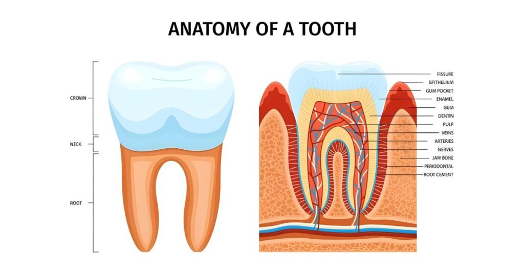
External Anatomy of a Tooth
Teeth include visible external structures that each serve specific purposes. The crown is the top part that protrudes above the gum line and is covered with enamel. It has a chewing surface designed to crush and grind food during mastication. The root anchors the tooth in the bone within the upper and lower jaw. It is encased by an outer layer called cementum. Between the crown and root is a narrowed region called the neck, which demarcates these two segments.
Enamel is the hardest substance in the human body and coats the outermost layer of the tooth crown. Its high mineral content, primarily composed of hydroxyapatite crystals, gives enamel its characteristic rigidity and strength. This protects the internal structures of the tooth from damage due to biting forces and exposure to substances in the oral environment. Enamel does not contain any living cells and is avascular, meaning no blood vessels run through it. However, it is attached to softer, living tissues beneath it via an interlocking system of rods and proteins that provide structural support.
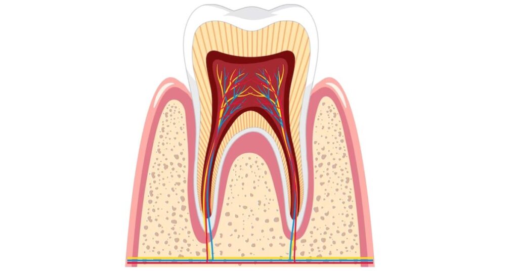
Internal Anatomy of a Tooth
Below the enamel is a thick middle layer of bonelike tissue called dentin. Dentin contains less mineral content than enamel so it is softer and more flexible, but it still protects the innermost structures of the tooth. Within the dentin are tiny tubules that run from the inner pulp to the dentin enamel junction near the outer enamel surface. These tubules house extensions of cells called odontoblasts that function in dentin formation. The tubules also contain fluid and microscopic nerve fibers, which is why damage to dentin can provoke tooth sensitivity.
At the center of a tooth is the dental pulp, which contains soft connective tissues, blood vessels, and nerves that enter through the root. The pulp nourishes the tooth and allows it to sense hot, cold, pressure, and pain. Odontoblasts that line the outer pulp chamber also aid in dentin production. Under normal conditions, the pulp is sterile due to the lack of a direct connection between it and the external environment. However, dental caries, chips or cracks in the enamel, or trauma can expose the pulp and put it at risk of infection. Protecting the pulp is paramount to maintaining tooth vitality and preventing disease.
Different Types of Teeth

Humans have four distinct types of teeth that work synergistically when biting and chewing:
Incisors
The front four teeth in both the upper and lower jaws are incisors. These have sharp, chisel-shaped crowns ideal for cutting and shearing food. Their enamel is thicker on the front surface than the back for added strength when incising.
Canines
The pointed canine teeth flank each side of the incisors. Their more pronounced cusps help puncture and tear food. Canines have the longest root of all tooth types to anchor them firmly in place. They serve a secondary purpose in humans – acting as guideposts to enable proper contact between the upper and lower teeth.
Premolars
Behind the canines are two premolars in each jaw quadrant. These have crowns with raised cusps that crush and tear harder foods. Their location provides excellent anchorage through two solid roots.
Molars
The most posterior teeth are broad, multi-cusped molars. Their expanded crowns and roots grind food into smaller particles to aid digestion. First molars have the largest surface area of any tooth to pulverize hard or fibrous foods.
The Importance of Understanding Tooth Anatomy for Oral Health

Appreciating the specialized design elements of different tooth types allows a better understanding of how to care for them. The thin enamel layer is vulnerable to acid erosion from sugary foods and beverages. Its lack of blood supply also means enamel cannot regenerate if lost to decay. Therefore, limiting sugar intake and maintaining diligent oral hygiene is critical to avoid permanent damage.
Knowing the composition of underlying dentin and pulp tissues explains why tooth sensitivity can occur when these regions are exposed. Prompt treatment of cavities and avoiding behaviors that wear away enamel, like aggressive brushing or grinding, help prevent this pain. Being aware that the pulp contains innervation and blood flow provides insight into why procedures like root canals are sometimes necessary for the resolution of infection and inflammation.
Overall, insight into tooth anatomy empowers individuals to make smart dietary choices and engage in proper hygiene techniques that maintain tooth integrity and function. It also enables comprehension of dental treatments so patients can make informed decisions about their oral health management. Just a short time invested in learning about these intricate body parts yields great dividends in terms of better appreciating and caring for our amazing teeth.
Conclusion
Our teeth are living wonders of biology, whose solid yet elegant structure enables them to withstand a lifetime of wear. Both the visible crown and hidden root regions have unique characteristics tailored to their purpose. Enamel, dentin, and dental pulp each contribute their properties to create a fully functional organ. The four types of teeth – incisors, canines, premolars, and molars – are strategically shaped to cut, tear, and grind food with maximum efficiency. Appreciating the anatomy of teeth allows for better oral hygiene practices and wiser dental health choices to keep these intricate structures in optimal condition.